Since the first Phenom desktop SEM devices developed during 2006-2007, this technology has undergone tremendous evolution. About two years ago, Thermo Fisher Scientific, the current owner of the Phenom product range, launched the sixth generation Phenom ProX G6.
Phenom Pharos G2: Desktop SEM with FEG resolution within the reach of every lab
Phenom Pharos G2: Desktop SEM with FEG resolution within the reach of every lab
The democratization of SEM technology
With the rise of the desktop SEM technology, electron microscopy has found access as an image analysis and analytical technique in thousands of labs worldwide. Not only the affordable investment budget and the low cost-of-ownership, but more importantly the “desktop” aspect (meaning the limited need for specialized infrastructure in terms of lab space, vibration reduction, control of ambient temperature, pressure, etc.) and the intuitive user interface (allowing the technology to be used by a large number of non-specialized operators) are responsible for this spectacular breakthrough. “The democratization of SEM technology” as one speaker called it during a speech at a recent congress.
A next step in the development of desktop SEM technology came with the integration of FEG-SEM technology into a desktop SEM. FEG-SEM (Field Emission Gun – Scanning Electron Microscope) provides the very highest resolution imaging compared to regular SEM. It guarantees high brightness, crisp images and a stable beam current.
Usually FEG-SEMs are large floor-standing systems, but the same high-resolution technology is now available with the Thermo Scientific Phenom Pharos.

Want to know more about the Phenom Pharos?
Download the Phenom Pharos product brochure here.
The highest resolution

Alumina coating
FEG gives the very highest resolution, with high brightness, crisp images and a stable beam current.
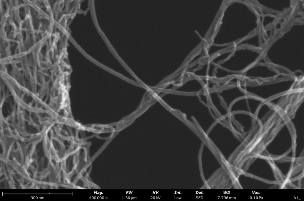
The Thermo Scientific Phenom Pharos G2 offers a resolution of ≤2nm @20kV SE. This shows the shape of nanoparticles, imperfections in coatings and other features that would be missed by other SEMs, including those with a traditional tungsten source.
Now in a desktop instrument
Field emission SEMs are usually large systems requiring a dedicated room with special infrastructure and connections. They’re often difficult to operate, requiring highly trained specialists. For this reason, FEG-SEM work is often outsourced to service labs or central facilities.
However, having a FEG-SEM in-house is now a much more realistic option. The Phenom Pharos G2 Desktop FEG-SEM is the only desktop SEM with a FEG source. It’s easy to install and operate, so you no longer need to rely on external services.
The Phenom Pharos has all the capabilities of a floor-standing FEG-SEM, housed in a tabletop model. It often outperforms floor-standing systems in terms of image quality, with significantly better user experience.
While other SEMs can end up being fully booked, the Phenom Pharos G2 performs imaging and analysis so quickly that in many facilities it’s used as a walk-up tool.

Phenom Pharos G2
Convenient and Easy to Use

Bacteria sample
With a small tabletop footprint, the Phenom Pharos G2 doesn’t take up much space, so it can be housed easily in your lab. You can then access results much faster than when using a floor-standing model housed elsewhere.
It is a reliable system, with robust parts and an integrated UPS to prevent power outage issues. Field emission tips generally cost more, but they are long-lasting. They usually last more than a year, so there’s less down-time for source exchange.
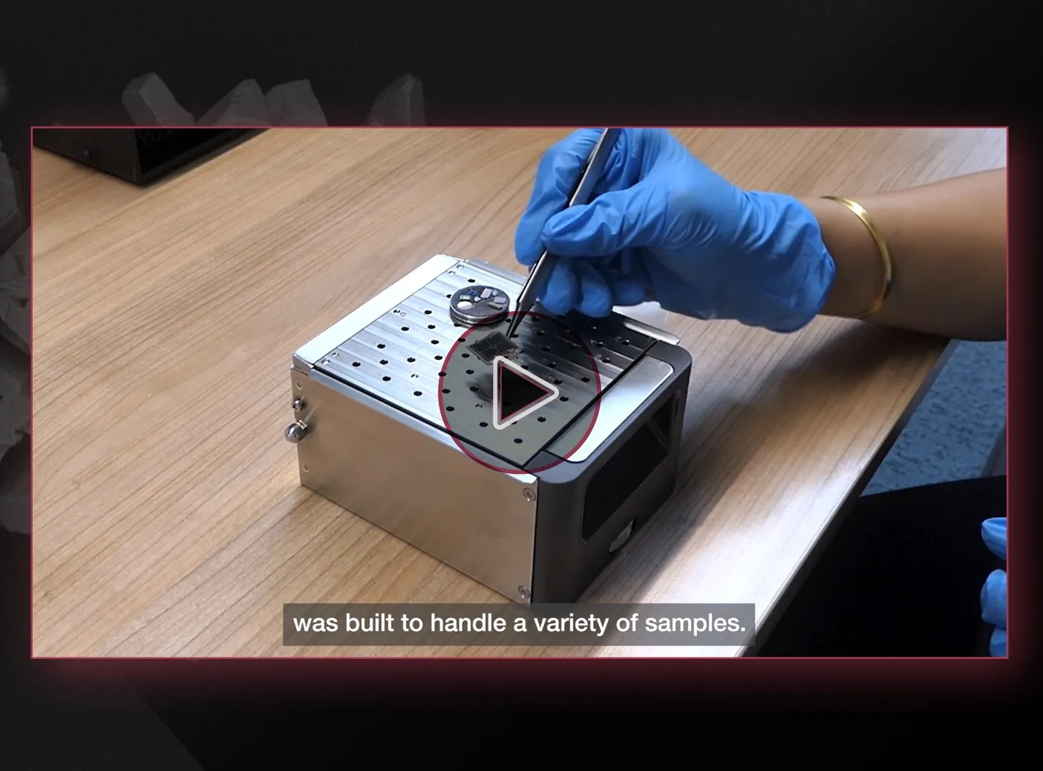
The Phenom Pharos is intuitive, easy-to-learn and highly productive, right from the start. Once a sample is loaded, an optical image is available immediately for navigation. After switching to SEM mode, an image appears in 30 seconds with an incredible amount of detail. It’s then easy to zoom in and navigate around the sample.
For demanding applications
While optical microscopes and tungsten SEMs provide high-resolution imaging, some applications require the higher resolution of a FEG-SEM, for example:
- Morphology of nanoparticles.
- Small defects in thin films.
- Insulating materials.
- Materials sensitive to high-kV electron beams.
A field emission SEM provides a stable, high-brightness beam for these demanding materials.

Carbon nano tubes
Soft Samples

Foam structure
Due to the high performance at low acceleration voltage of FEG compared to other SEM electron sources, you can study insulating and beam-sensitive materials without sample preparation. Samples are not damaged by the beam and nanoscale features are clearly visible.
The expanded energy range gives you the ability to image beam-sensitive specimens such as:
- Zeolites – used in water purification, pharmaceutical powders and many common tablet and capsule-based medicines.
- Polymer fibers – in materials such as nylon, polyester, spandex and Kevlar.
Low-kV imaging is essential for studying these types of materials, because higher energy electrons damage their structure.

Request a demo
Discover superior imaging with Phenom Pharos’ advanced field emission gun technology and versatile voltage range. Request a demo or more information now!


